- Conformation Analysis
- Protein Structure Alignment
- Protein Misfolding vs. Alzheimer Disease
- Fragment Search
- Evaluate Structural Similarity for Proteins with Diverse Degrees in Homology
- Mutation Analysis
- Analysis of CDR Loops in Antibody
- Comparison of Insulin Receptor vs. IGF-1 Receptor
- Drug Binding Site
- Predicting Off Target Hits in Drug Discovery
-------------------------------------------------------------------------------------------------------------------------------------------------------------
Drug Binding Site
ATP Binding Sites on Protein Kinases
This sample shows that same inhibitor can bind different kinases; same kinase binding site can accept different molecules.
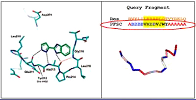
- Crystal structure of pyrazole-benzimidazole fragment complexes with Aurora A. (PDB ID = 2W1G)
- ATP Binding sites on protein kinases is described by PFSC
-
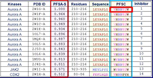
With PBSDD technology, 12 targets was found with similar binding sites after screening the kinases library of protein structures. The PFSC displays the folding conformations.
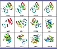
- Images of three-dimensional structures of 12 protein kinases from Protein Databank (PDB). 12 structures belong to different kinase groups, and each Kinses bind to different inhibitors. Aurora A: 2W1G, 2W1F, 2W1D, 2W1C, 2W1E, 1OL7, 1MQ4, 1OL5, 2J50 and 1OL6; JAK2: 2W1I; CDK2: 2W1H.
- 12 binding site images, core residues, sequences and PFSC of 12 ATP binding sites. 12 structures belong to different kinase groups, and each Kinase bind to different inhibitors. Aurora A: 2W1G, 2W1F, 2W1D, 2W1C, 2W1E, 1OL7, 1MQ4, 1OL5, 2J50 and 1OL6; JAK2: 2W1I; CDK2: 2W1H
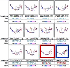
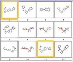
- 12 ATP inhibitors images. Each inhibitors exists on perspective ATP binding site on left figure. Compound 1-10 binds to Aurora A: 2W1G, 2W1F, 2W1D, 2W1C, 2W1E, 1OL7, 1MQ4, 1OL5, 2J50 and 1OL6; JAK2: 2W1I; CDK2: 2W1H.
Reference
- Howard, S., Berdini, etc., Fragment-Based Discovery of the Pyrazol-4-Yl Urea (at9283), a Multitargeted Kinase Inhibitor with Potent Aurora Kinase Activity (Dagger). (2009), J.Med.Chem. 52: 379-388